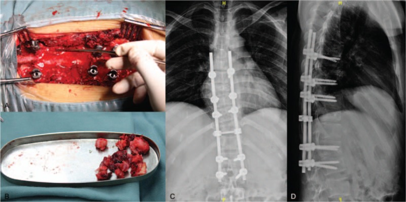
Figure 3.

A, Intraoperative photography depicting the exposed spinal cord. B, Intraoperative photography depicting partially resected metastatic tumor. C, Posteroanterior (PA) x-ray image of the thoracic spine obtained postoperatively. D, Lateral x-ray image of the thoracic spine obtained postoperatively.
