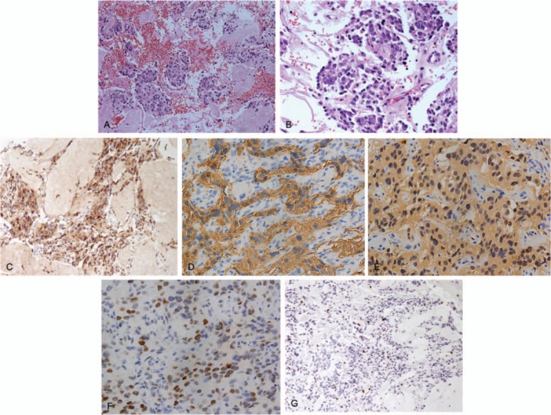
Figure 4.

Pathologic histology of spinal metastases. A and B, Microphotography showing characteristic nests of tumor cells separated by vascular septa (Zellballen) with cells showing significant nuclear pleomorphism with prominent nucleoli (Hematoxylin and Eeosin, original magnification ×20 and ×40). C, Chromogranin A immunostaining is strongly positive in the chromaffin cells. Chromogranin A is present in the secretory granules. D, Synaptophysin immunostaining shows strong, diffuse cytoplasmic staining in the tumor cells. E, The sustentacular cells of the spinal metastases of pheochromocytoma showing characteristic staining of S100. F, P-53 immunostaining is sporadically positive. G, Ki-67 immunostaining shows 3% Ki-67 positive cells. Ki-67 staining is localized in the tumor nuclei.
