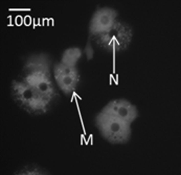
Fig. 2.

Representative ×40 magnification of primary hepatocytes stained with TMRM mitochondrial stain. Mitochondria (M) are indicated by red punctae and an example shown using the arrow labelled M. The nucleus (N) of the hepatocytes in indicated using the arrow labelled N. The scale bar represents 100 µm. (Color figure online)
