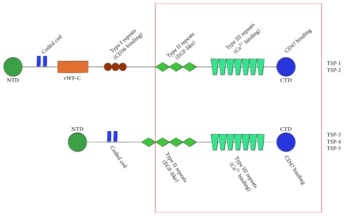
Figure 1.
Schematic representation of TSPs. All of TSPs share highly homologous CTD, Type 2 repeats, and Type 3 repeats (red region), while TSP-1 and TSP-2 have vWF-C domain and Type 1 repeats. NTD may be characteristic to the family members. TSPs have a complex multidomain architecture that provides an option to bind various ligands. For instance, CTD is involved in CD47 binding, while Type III repeats contain Ca2+ binding site. Type I repeats are implicated in interaction with CD36, a receptor for TSP1 and TSP2, and inhibition of MMPs, while vWF-C is responsible for binding members of the TGF-β superfamily. This figure is only a partial listing. CTD: C-terminal domain, EGF-like: epidermal growth factor-like, vWF-C: von Willebrand factor C-type, and NTD: N-terminal domain.

