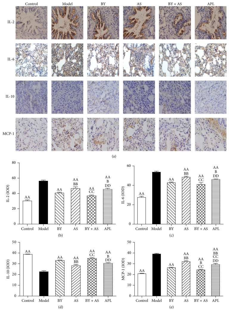
Figure 4.
Changes of inflammatory cytokines in the lung in all treatment groups. Immunohistochemical staining of lung sections (magnification, ×400) (a); IL-2, IL-6, IL-10, and MCP-1 were quantitatively analyzed (b, c, d, and e). Values are expressed as the mean ± SEM. AAP < 0.01 versus Model group; BBP < 0.01, BP < 0.05 versus BY group; CCP < 0.01 versus AS group; DDP < 0.01 versus BY + AS group.

