Fig. 2.
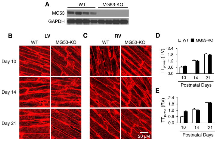
Deficiency of MG53 does not alter T-tubule maturation. A, Immunoblots of MG53 protein expression in heart lysates from WT and MG53-KO mice. GAPDH: loading control. B & C, Representative confocal of left ventricular (LV, B) or right ventricular (RV, C) T-tubules from WT or MG53-KO mice at postnatal days 10, 14, and 21. T-tubules were stained with a lipophilic marker MM 4–64. D & E, Mean values of TTpower in the LV (D) and RV (E) of WT and MG53-KO mice at different ages. N=4 mice in each group. Data shown are mean ± SE.

