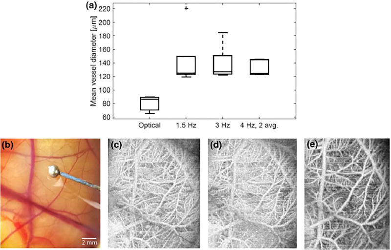
FIGURE 2.
(a) The mean vessel diameter was measured optically and with acoustic angiography in 12 animals, yielding mean values of 65 lm (optical) and 119 lm (acoustic angiography). (b) Illustrative optical image of the CAM and (c) matched maximum intensity projection in the same embryo at acquisition rates of 1.5, 3, and 4 frames per second (2 frame averaging) show that different vessels are detected with different acquisition rates. The diameter of ball bearing (reference object) in (a) is 1.60 mm.

