Fig. 3.
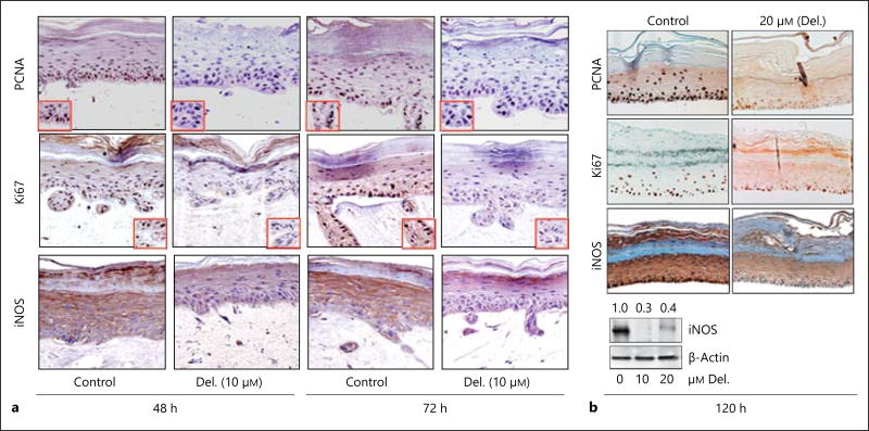
Delphinidin (Del.) treatment reduces the protein expression of proliferation (PCNA and Ki67) and inflammation (iNOS) markers in 3D reconstituted human PSE. Representative photomicrographs of immunohistochemical staining showing that proliferation and inflammation markers are strongly and predominantly expressed in the basal and granular layers of control PSE. a More intense staining of PCNA, Ki67 and iNOS protein expression was observed in control PSE tissue sections, which was markedly depressed but not completely absent in 48- and 72-hour delphinidin-treated tissues (10 µM) compared to untreated controls. b Protein expression of PCNA, Ki67 and iNOS were markedly and almost completely inhibited in delphinidin-treated constructs (20 µM) after prolonged treatment of 120 h. Immunoblots of iNOS (lower panel) expression in control vs. delphinidin-treated PSEs. Bars = 20 µm.

