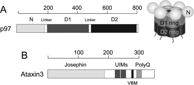
Figure 1.

Schematic representation of p97 and ataxin3. A, structure of p97. At left is shown the domain organization of the p97 protomer. Each protomer comprises an N-terminal domain shown in light gray, D1- and D2-ATPase domains shown in dark gray and black, respectively, and an unstructured C-terminal region. At right is a schematic of the assembled hexamer, using the same shading. The D1- and D2-domains form two coaxially-stacked rings around a central pore, with the N-domains arranged along the periphery of the D1 ring. B, domain organization of ataxin3 showing the Josephin domain, two ubiquitin-interacting motifs (UIMs), the p97/VCP-binding motif (VBM), and the polyglutamine (polyQ) repeat region. The linkers, UIMs, VBM, and polyQ regions are not drawn to scale. The scales above each domain representation show length in amino acids.
