Figure 4.
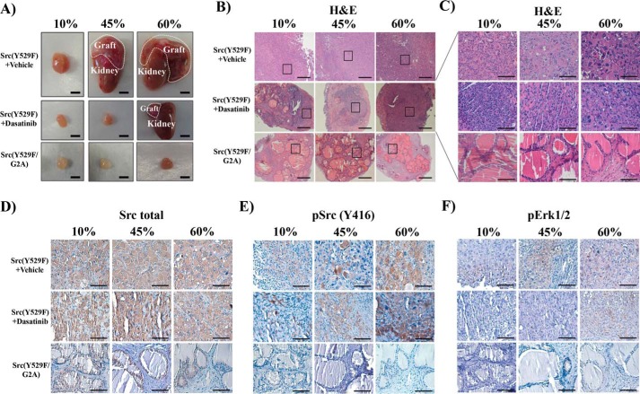
Loss of myristoylation inhibits Src-mediated HFD-accelerated tumor progression. A, the in vivo prostate regeneration assay was performed with Src(Y529F) and Src(Y529F/G2A) under 10%, 45%, and 60% fat diets with vehicle or dasatinib treatment (75 mg/kg/day weeks 5 to 8), a similar experimental setting as described in Fig. 1. The dashed lines represent the regenerated prostate tumors grown on the kidney (scale bars = 4 mm). B–F, H&E panoramic view (B, scale bars = 300 μm) and selected region (C, scale bars = 100 μm) and IHC staining of total Src kinase, pSrc(Tyr-416), and pErk1/2 of regenerated prostate tissue (D–F, scale bars = 100 μm) in A. See also supplemental Fig. S5.

