Figure 5.
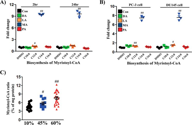
Exogenous MA or an HFD increases intracellular myristoyl-CoA in cells or xenograft tumors. A, 293T+TRE/Src(Y529F) cells were treated with DMSO, DA (C10:0), LA (C12:0), MA (C14:0), or PA (C16:0) for 2 or 24 h (three repeats in each group). Con, control. B, PC-3 or DU145 cells were treated similarly for 24 h. The levels of myristoyl-CoA were analyzed by LC/MS-MS, and the levels in the control were set as 1 (three repeats in each group). C, the levels of myristoyl-CoA in PC-3 xenograft tumors from Fig. 1 were analyzed by LC/MS-MS. The amount of myristoyl-CoA was standardized to the amount of total protein in the tumors. Sixteen tumors per group were analyzed. #, p < 0.05; ##, p < 0.01. See also supplemental Fig. S6.

