Figure 3.
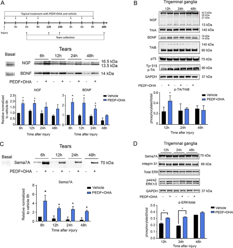
PEDF + DHA treatment increases secretion of NGF, BDNF, and Sema7A in tears and the phosphorylation of TrkB and ERK1/2 in TG. A, corneas were injured and treated with PEDF + DHA and tears collected as shown in the experimental design (top). Seven micrograms of protein from the mouse tear film (pool of six eyes/sample) were used for Western blot analysis of BDNF and NGF (bottom). B, Western blot analysis of TrkA, TrkB, p75, Tyr-phosphorylated Trks, and GAPDH in the TG (pool of six TGs, 50 μg of protein per well). Bars in A and B represent the mean of two experiments (two different pooled sample sets for each experiment, four samples in total) ± S.D. *, p < 0.05 with the t test analysis in comparison with the vehicle at the same time point. C, Western blot of Sema7A secreted to tears after PEDF + DHA treatment. Seven micrograms of protein collected from tears (pool of six eyes/sample) were used. D, Western blot analysis of intracellular integrin β1, total ERK, p44/p42 ERK1/2, and GAPDH in the TG (pool of six TGs, 50 μg of protein per well) of corneas treated with PEDF + DHA or vehicle. Details about the antibodies used are in Table 3. Bars in C and D represent the mean of two experiments (two different pooled sample sets for each experiment, four samples in total) ± S.D. of p-ERK/total ERK ratio. *, p < 0.05 with t test analysis in comparison with the vehicle at the same time points.

