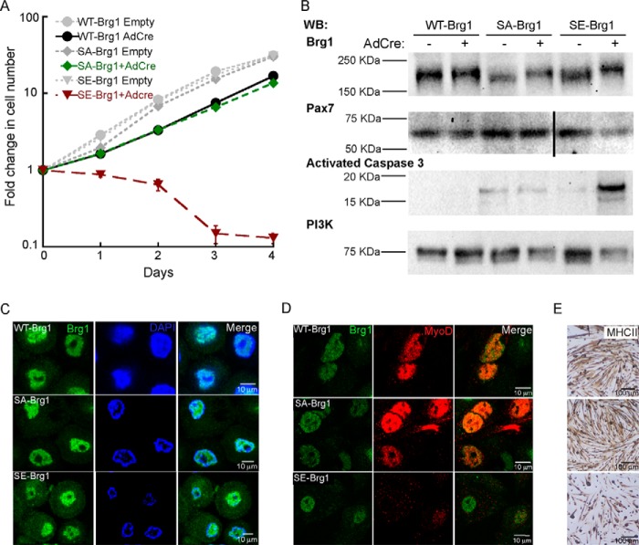
Figure 3.
Phosphomimetic mutations in Brg1 inhibit proliferation and reduce viability of primary myoblasts. A, proliferation assay of Brg1-deficient (Brg1 c/c) cells transduced with WT-, SA-, or SE-Brg1. Data represent the average of three independent experiments ± S.D. B, representative Western blots (WB) showing the expression of Brg1, Pax7, activated caspase 3, and PI3K as a loading control. C, representative confocal microscopy images showing nuclear localization of WT-, SA-, and SE-Brg1 expressed in proliferating primary myoblasts. D, representative confocal microscopy images of primary myoblasts transduced with the WT-, SA-, and SE-Brg1 mutants that were differentiated for 48 h and immunostained for Brg1 and MyoD. E, representative light microscopy images of the phenotypes observed when myoblasts expressing the indicated Brg1 protein were differentiated for 48 h; cells were immunostained using an anti-myosin heavy chain II (MHCII) antibody.

