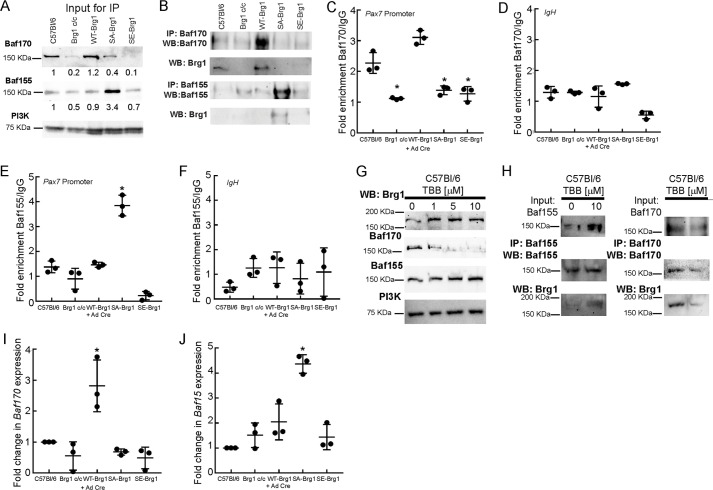
Figure 7.
Inhibition of CK2 or expression of phosphomimetic or non-phosphorylatable Brg1 mutations determines interaction with Baf170 or Baf155. A, representative Western blots (WB) showing 12% of the input for the subsequent immunoprecipitation (IP) experiments, which documents expression of Baf170 and Baf155 in the indicated proliferating primary myoblasts. Bands were quantified using ImageJ. Brg1 expression was normalized to PI3K expression, and the value of expression in the C57Bl/6 myoblasts was set at 1. Relative expression levels are indicated below each band. B, representative profiles of Baf170 and Baf155 immunoprecipitations of Brg1 from the indicated primary myoblasts. C and D, ChIP experiments showing Baf170 recruitment to the Pax7 promoter (C) or to the IgH enhancer (D) when endogenous or WT-Brg1 is expressed. E and F, ChIP experiments showing Baf155 recruitment to the Pax7 promoter (E) or to the IgH enhancer (F) when the non-phosphorylatable Brg1 mutant is expressed. Data in C–F represent the average of three independent experiments, each assayed in triplicate ± S.D.; *, p < 0.01. G, representative Western blottings showing the expression of Baf170, Baf15, and Brg1 in C57Bl/6 primary myoblasts treated with increasing concentrations of TBB. PI3K levels were measured as a control. H, representative immunoprecipitations of Baf155/Brg1 and Baf170/Brg1 from primary myoblasts treated with 10 μm TBB. I and J, relative levels of Baf170 and Baf155 mRNA present in the indicated myoblasts. Levels in C57Bl6 primary myoblasts were normalized to 1. Data represent the average of three independent experiments, each assayed in triplicate ± S.D.; *, p < 0.01.

