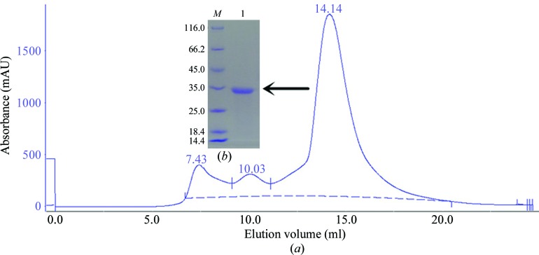
Figure 1.
Purification of PhyH-DI using a gel-filtration column and SDS–PAGE analysis. (a) Purification profile of PhyH-DI, which eluted as a symmetrical peak from a Superdex G200 SEC column. (b) 15% SDS–PAGE stained with Coomassie Brilliant Blue. Lane M, protein marker (labelled in kDa); lane 1, PhyH-DI corresponding to the peak on the gel-filtration profile (0.5 mg ml−1).

