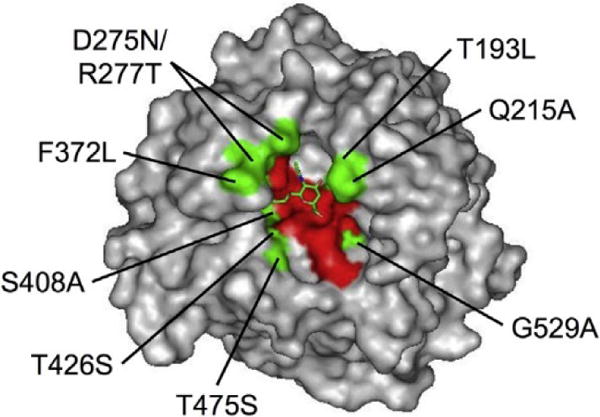
Fig. 1.

Locations of amino acid substitutions and the sialic acid binding pocket in the HN3 glycoprotein. The three-dimensional structure of HN3 glycoprotein with sialic acid (PDB ID: 1V3C) was shown by PyMOL software. There were 9 amino acid residues that were different in sequences of the HN1 and HN3 glycoproteins in or neighboring the sialic acid binding pocket (green residues). These 9 residues of the HN3 glycoprotein (left of position numbers) were substituted for the corresponding residues of the HN1 glycoprotein (right of position numbers). Red residues mean that amino acids directly interacted with the sialic acid residue through a hydrogen bond. Sialic acid is shown by a stick model in green.
