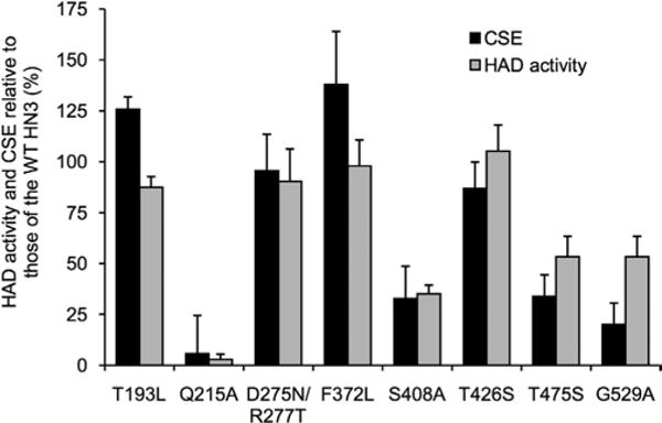
Fig. 2.

HAD activity and CSE of amino acid-substituted HN3 glycoproteins. COS-7 cells were transfected with HN3 genes. CSE (filled column) was quantitated by flow cytometry with rabbit anti-hPIV3 antibody. HAD activity (gray column) was determined by the amount of native RBCs adsorbed on the HN3 glycoprotein-expressing cells. All data are represented as relative percentages to the WT HN3 glycoprotein.
