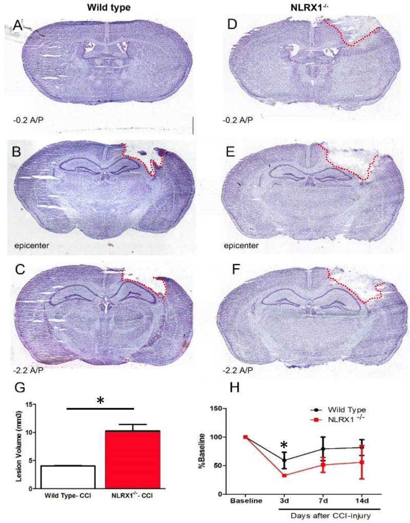
Figure 1. Increased lesion volume and motor deficits in Nlrx1−/− mice following CCI-injury.
A–C) Nissl staining of wild type brains at 3 days post-CCI compared to Nlrx1−/−(D–F) shows increased lesion volume (G) in the absence of NLRX1. (H) Motor deficits are also more prominent in Nlrx1−/− mice compared to wild type at 3–14 days post-CCI. (n=5–7 per group; **p<0.05).

