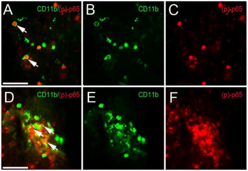
Figure 4. Microglia expression of (p)-p65 at 3 days post-CCI injury in the cortex of wild type and Nlrx1−/− mice.
(A–C) High magnification fluorescence image of CD11b (green) and phospho(p)-p65 (red) in wild type injured cortex. (D–F) Increased CD11b/ phospho(p)-p65-positive cells are seen Nlrx1−/− mice. Scale bar = 100µm in images A and D.

