
Figure 3.
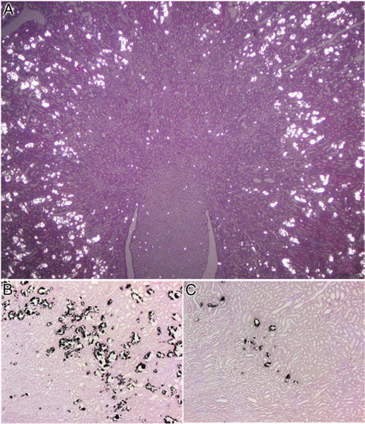
Light microscopy. A, rat kidney with significant CaOx crystal deposition as seen under polarized light. H & E, reduced from ×1.2. B, in kidney section crystals appear as dark deposits. Pizzalato stain, reduced from ×10. C, kidney of hyperoxaluric rat treated with apocynin demonstrates highly significant decrease in CaOx crystal deposition. Reduced from ×10.