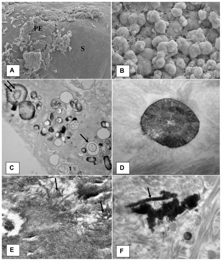
Figure 2.
Microscopic appearance of the Randall’s plaque. A). Plaque appears as a bulge on the papillary surface. Epithelium (PE) is eroding exposing sub-epithelial amorphous layer (S). B). Spherical deposits of CaP mixed with fibrous material are present under the papillary surface epithelium. C). Calcification consisting of large and small vesicles (arrows), some with crystalline material inside (double arrow) are present embedded in an amorphous to granular to fibrous substrate. D). Concentrically laminated spherical unit surrounded by fibrous substrate. E). Calcification consisting of large dark deposits. Center of the deposit showing plate-like individual crystals (arrows). F). Calcification surrounded by what appear to be collagen fibers with distinct banding pattern (arrow). Arrow points to a calcified collagen fiber showing beaded exterior. Adapted from Khan and Canales, 2015, Urolithiasis 43 Supplement 1:109–123 (10).

