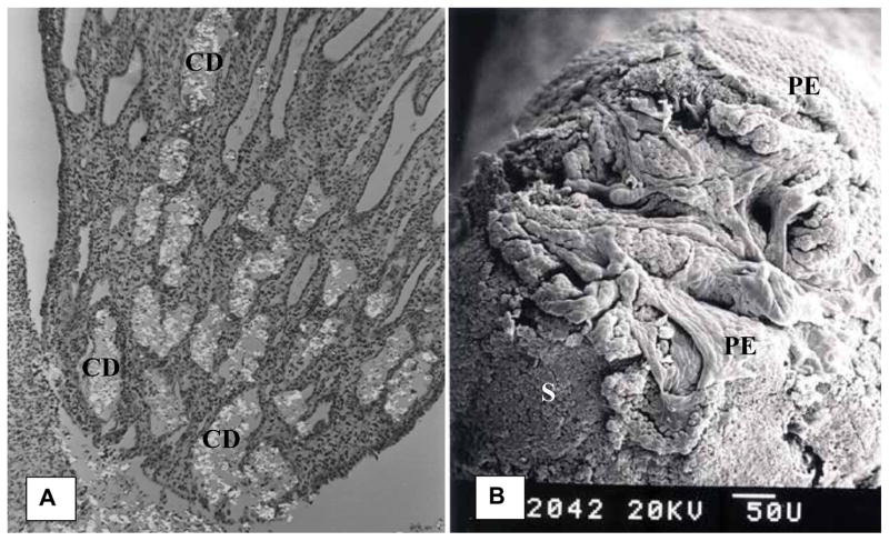
Figure 9.
Papillary tip of a rat with hyperoxaluria. A). Light microscopic appearance of a longitudinal section showing birefringent CaOx monohydrate crystals plugging the collecting ducts (CD). B). Scanning microscopy of the papillary tip. Surface epithelium (PE) is eroding exposing CaOx deposit (S) plugging the ducts of Bellini. Openings of the ducts appear impaired.

