
Figure 6.
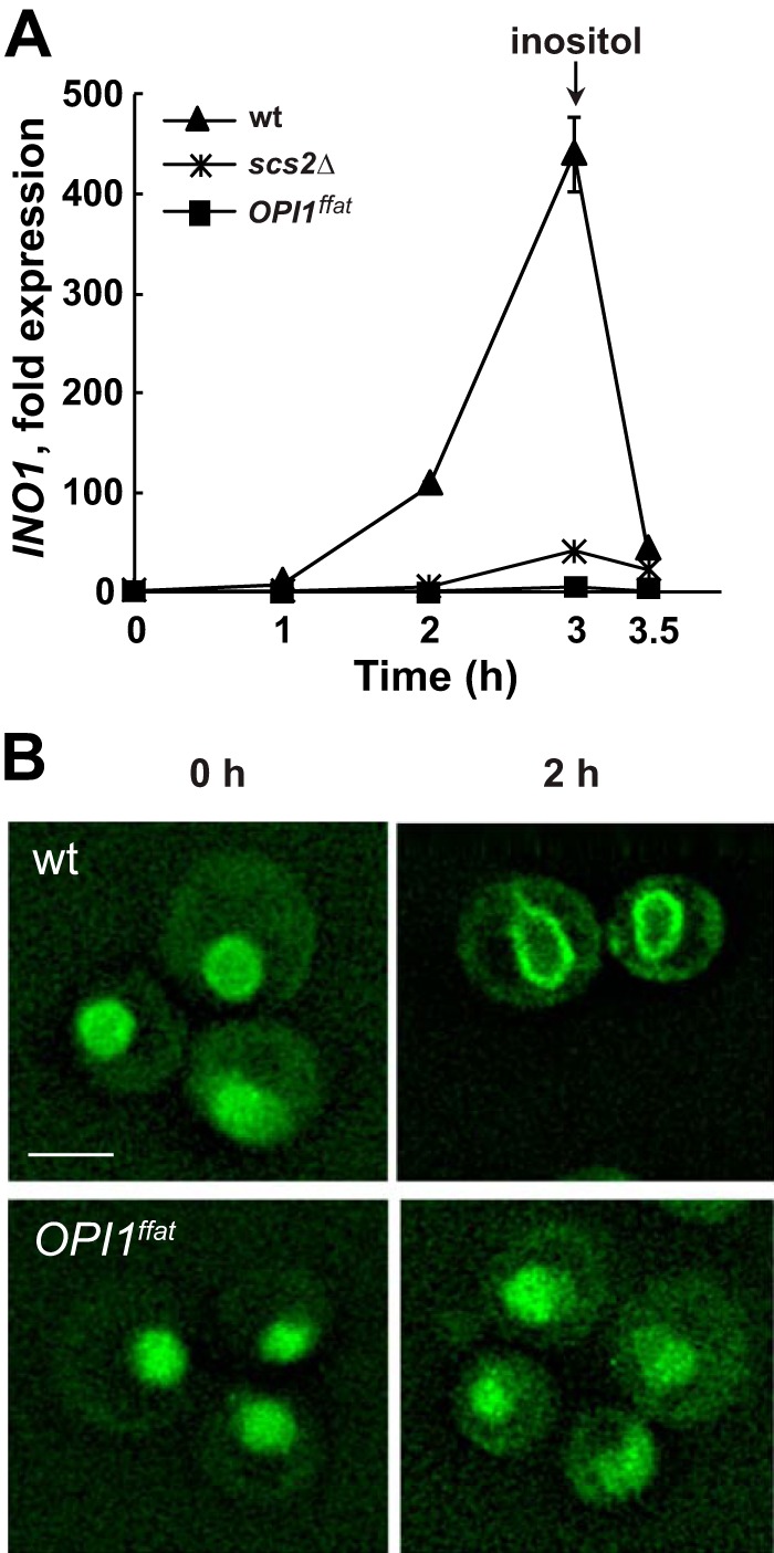
INO1 expression in wild-type, OPI1ffat, and scs2Δ strains and localization of Opi1p-GFP in wild-type or Opi1ffatp-GFP in scs2Δ and OPI1ffat strains, respectively, after a shift to I−C+ medium. A, overnight cultures of YCY3 (wild type), YCY5 (OPI1ffat), and YCY7 (scs2Δ) expressing genomic Opi1p-GFP or Opi1pffat-GFP, respectively, were diluted to A600 = 0.2 in I+C+ medium and allowed to grow to mid-logarithmic phase at 30 °C. Cells were harvested by centrifugation, washed and resuspended in I−C+ medium, and incubated for 3 h. Inositol was added back after 3 h of inositol starvation. Samples were taken at 0, 1, 2, and 3 h of inositol starvation and 30 min after adding back inositol. Total RNA was isolated and analyzed by RT-PCR as described under “Experimental procedures.” Solid triangles, wild type; solid squares, OPI1ffat; solid crosses, scs2Δ. B, Opi1p-GFP localization in wild type and Opi1pffat-GFP in OPI1ffat at 0 and 2 h after a shift to I−C+ medium. Cells were imaged by fluorescence microscopy. A representative z-section was chosen for each image. Scale bar, 5 μm.