Fig. 3.

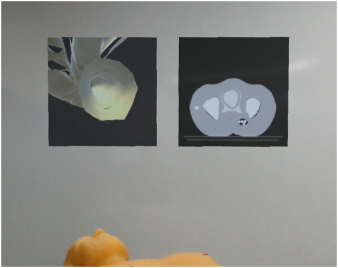
Two virtual canvasses are hovering above the patient. The left canvas shows the PoV of the catheter inside the vessel and the right canvas renders the representative CT slice in respect to the tracked catheter position

Two virtual canvasses are hovering above the patient. The left canvas shows the PoV of the catheter inside the vessel and the right canvas renders the representative CT slice in respect to the tracked catheter position