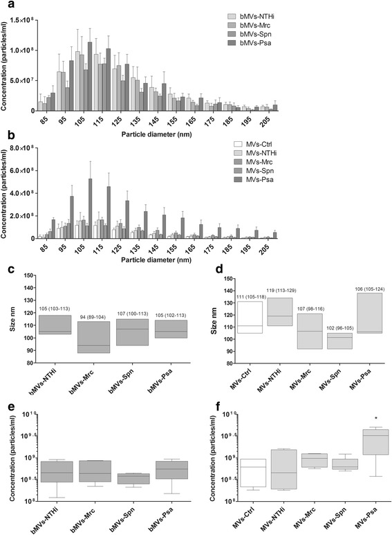
Fig. 2.

Tunable resistive pulse sensing-based characterization of MVs released upon infection with or culture of NTHi, Mrc, Spn, and Psa. Conditioned media of bacterial cultures (bMVs) were used to assess the vesicle size and numbers of vesicles released (a). Also, MV release by uninfected THP1 macrophages (MVs-Ctrl) and by THP1 macrophages after 4 h of infection with different bacteria (MVs-NTHi, -Mrc, -Psa, -Spn) was determined (b). The median diameter and interquartile range (between brackets) was calculated and is given for bacterial MVs (c) and for the mixed MV population released by cells and bacteria during infection (d). Box and whisker plots (e-b) indicate the median (line in box), 25th and 75th percentiles (outer lines of box) and the minimal and maximal values (whiskers) for the bacterial MV concentration (e) and the concentration of MVs released upon infection (f). Results are from 6 independent experiments. *p < 0.05
