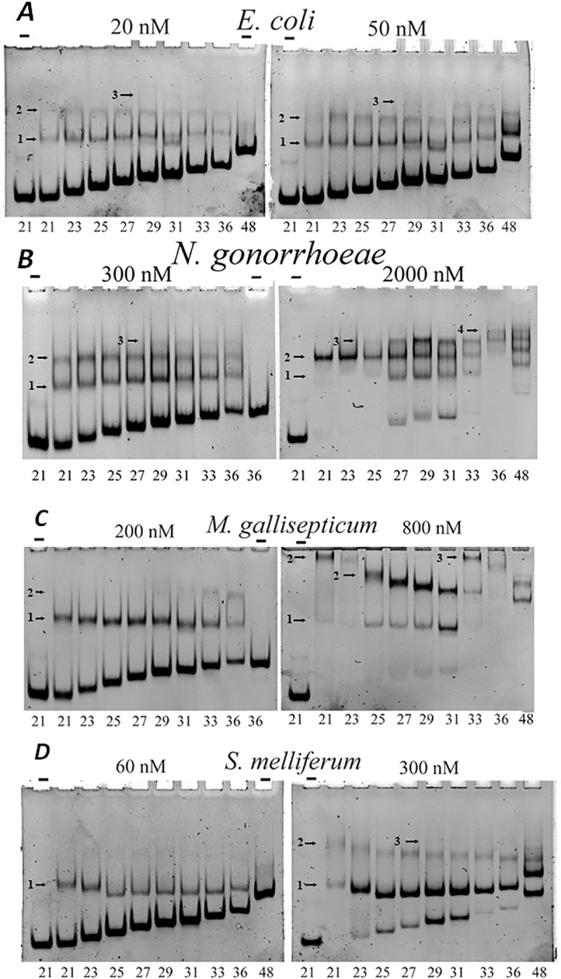
Fig 9. HU binding to dsDNA of various lengths.

Binding of labeled DNA to HU proteins was analyzed by polyacrylamide gel electrophoresis. The gel was buffered with 50 mM Tris–borate; binding mixture contains 40 mM NaCl. DNA samples were: dsDNA of sequence ‘D’ with the length varying from 21 to 48 bp (indicated at the bottom). HU origin and concentration is indicated at the top (“-“, no HU was added). Bands corresponding to HU-DNA complexes are marked with arrows, the number of HU dimers in each complex is indicated on the left of the arrow. Panels correspond to HU proteins of various bacteria, protein concentrations are indicated.
