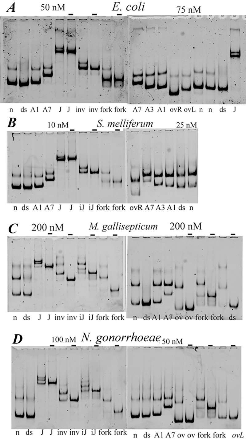
Fig 10. HU binding to “distorted” DNA structures checked by polyacrylamide gel mobility assay.

HU protein at concentrations indicated above the gel image (“-“, no HU was added) was mixed with 5’-labelled DNA in a buffer containing 150 mM NaCl; the bound and free DNA were gel-separated. DNA structures indicated at the bottom of the gel images: n, nicked DNA; ds, dsDNA; A1, A3 and A7, DNA bulges, containing one, three or seven non-paired adenines in one of DNA strands; J–four-way junction; fork, ssDNA fork; ov, DNA overhang; iJ, incomplete junction lacking one DNA strand; inv, DNA invasion. Panels correspond to HU proteins of various bacteria.
