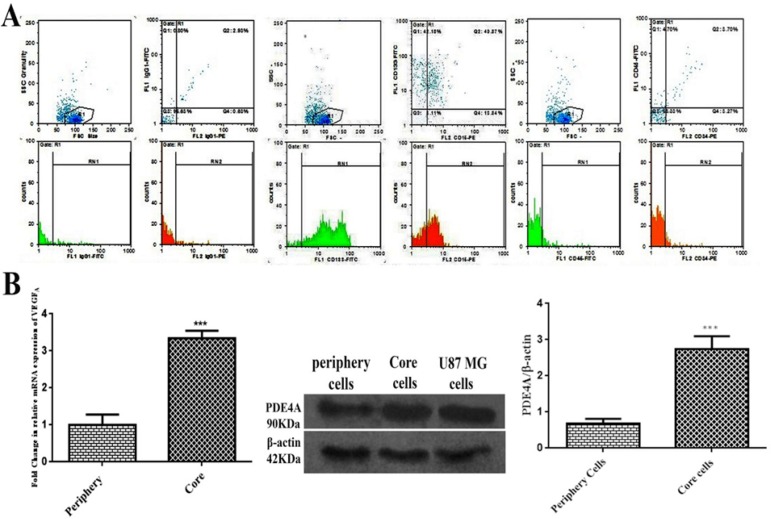
Figure 1.
Characterization of TICs isolated from GBM tissue. (A) Flow cytometry results of GBM-derived neurospheres in primary culture. TICs were positive for CD133 and CD15 but did not considerably express CD34 and CD45. (B) The relative expression of VEGFA mRNA measured by qPCR and immunoreactive bands and relative expression of PDE4A detected by western blot in the periphery-derived cells and core-derived TICs. U87 MG cells express PDE4A protein. Beta-actin was considered as housekeeping. Bars represent fold differences of mean normalized expression values±SEM (n=3). ***P<0.001 compared to periphery zone.

