
Figure 1.
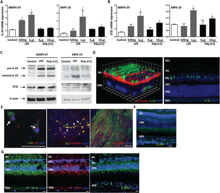
IL‐33 is expressed in murine and human retinas, and is induced in RPE. Expression of IL‐33 (A) and its receptor, ST2L (B), mRNA expression in retinal pigment epithelium (RPE) treated with TLR agonists [LPS, poly(I:C)] as indicated for 24 h (n = 4 per group). (C) Western blot analysis of IL‐33 and ST2 in mouse (B6RPE‐07) and human (ARPE‐19) RPE cells upon activation with TLR agonists. (D) Immunofluorescence staining showing expression of IL‐33 in adult murine retina. GCL = ganglion cell layer; IPL = inner plexiform layer; INL = inner nuclear layer; OPL = outer plexiform layer; ONL = outer nuclear layer. Scale bar = 100 µm. (E) ST2 expression in choroidal mast cells (arrows), choroidal fibroblasts (arrowheads), and RPE. (F) Immunofluorescence staining showing expression of IL‐1 receptor accessory protein (IL1RAP) in murine retina. Scale bar =100 µm. (G) Representative images of immunofluorescence staining showing expression of IL‐33, ST2, and IL1RAP in sections of human retina. Scale bar = 100 µm. Data are shown as mean ± SEM. Data are representative of at least three independent experiments with similar results. *p < 0.05. Statistical analysis was performed with Student's t‐test.