Figure 7.

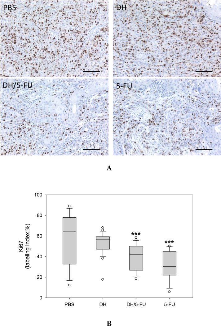
IHC staining of Ki67, a marker of proliferating cells in the tumor sections. (A) Representative images. Scale bar: 100 μm. (B) Fractions of Ki67-positive cells. *** p<0.001 vs. the PBS group.

IHC staining of Ki67, a marker of proliferating cells in the tumor sections. (A) Representative images. Scale bar: 100 μm. (B) Fractions of Ki67-positive cells. *** p<0.001 vs. the PBS group.