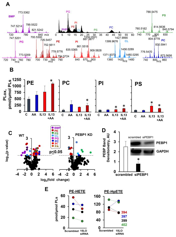
Figure 3. 15LO1 catalyzes PEBP1-dependent production of PEox in IL13 stimulated HAECs.
(A) Normal phase LC/MS/MS chromatogram (black) and mass spectra of PLs in HAECs. BMP-bis-monoacylglycero-phosphate; PG-phosphatidylglycerol; PE-phosphatidylethanolamine; PS-phosphatidylserine; CL-cardiolipin; PC-phosphatidylcholine; PI-phosphatidylinositol.
(B) Contents of PLox in HAECs treated with IL13 and AA (means±SD. *p<0.05 vs. control, N=3/group).
(C) Volcano plots of IL13 induced changes of PEox in wt (left plot) and PEBP1 KD HAECs (right plot) (log2 (fold-change) vs. significance (log10 p-value). Cells were exposed to IL13 in the presence of AA.
(D) Quantitation of PEBP1 KD by siRNA in HAECs (means±SD, *p<0.05 scrambled vs. siPEBP1, N=3/group). Insert: A typical Western blot for PEBP1.
(E) 15LO1 KD changes HETE-PE and HpETE-PE in HAECs exposed to IL13 and AA, (N=4, different human donors). See also Figure S3 and S4.

