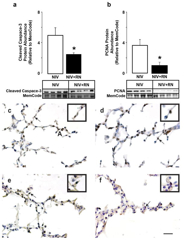
Figure 2.
Quantification of cleaved caspase-3 and PCNA protein abundance in preterm lambs supported by NIS, with or without restricted nutrition (RN). White fill indicates NIS control, black fill indicates NIS+RN. Results are shown as mean ± SD for n=4/group. Panel a: Cleaved caspase-3 protein abundance. Panel b: PCNA protein abundance. Cleaved caspase-3 and PCNA protein abundance are significantly lower in lungs of NIS+RN group compared to NIS control group (*different from the NIS group by Mann-Whitney U test, p< 0.05). Panels c and d: NIS immunohistochemistry of (c) cleaved caspase 3 and (d) PCNA. Panels e and f: NIS+RN immunohistochemistry of (e) cleaved caspase 3 and (f) PCNA. Immunohistochemistry shows that fewer mesenchymal-cell nuclei are immunostained brown for cleaved caspase 3 protein in NIS+RN and PCNA protein for the NIS+RN group compared to the NIS control group (scale bar is 100 μm). Insets are enlargements of the region identified by the arrow in each panel.

