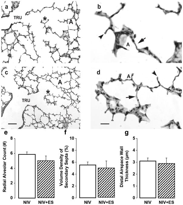
Figure 3.
Histologic appearance and morphometric quantification of lung structure in preterm lambs supported by NIS, with or without excess sedation (ES). Panels a and b: NIS control lung. Panels c and d: NIS+ES lung. TRU = terminal respiratory unit, DAS = distal airspace, A = alveolus. * identifies the enlarged region in panels b and d. Lung parenchyma of the NIS control group is similar to the corresponding NIS+ES group (panels a and c, the scale bar is 20 μm; panels b and d, the scale bar is 100 μm). Images are representative of the morphometric results shown in panels e, f, and g. Morphometric results are shown as mean ± SD. White fill indicates NIS control, hatched fill indicates NIS+ES. Radial alveolar count (panel e), volume density of secondary septa (panel f), and distal airspace wall thickness (panel g) is not different between NIS control group and NIS+ES group.

