
Figure 4.
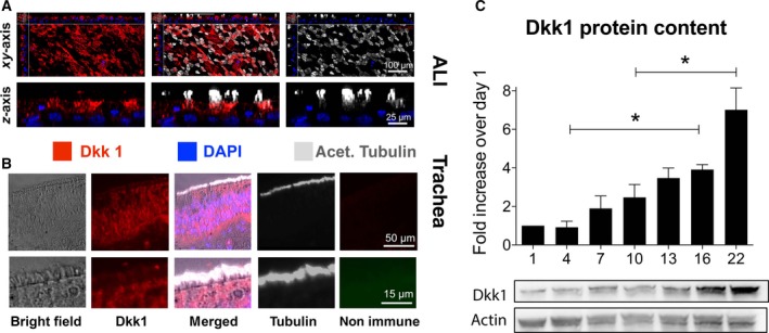
Dkk1 expression in human airway epithelial cells. (A) Confocal micrographs of differentiated ALI cultures labeled for Dkk1 (red), cilia (white) and nuclei (blue) showed Dkk1 expression. All images, but particularly z stacks, suggest localization of Dkk1 to ciliated cells. (B) In human tracheal sections from a normal individual, immunofluorescence showed the presence of Dkk1 in the airway epithelium, with expression in ciliated cells. (C) Quantification of Dkk1 protein by western blotting (normalized to actin) during differentiation of NHBE cells grown at the ALI showed a low Dkk1 expression in undifferentiated cells during the proliferative phase. A significant increase occurred during the differentiation phase using one‐way ANOVA and Tukey's multiple comparison test (*P < 0.05).