
FIGURE 1.
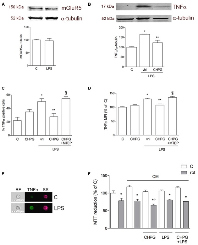
Metabotropic glutamate (mGlu5) receptor mediates anti-inflammatory effect on BV2 microglial cells. Western blot analysis of expression of mGlu5 receptor in BV2 cells treated with LPS (0.1 μg/ml) for 24 h (A). BV2 cells were activated with LPS (0.1 μg/ml for 6 h) after pretreatment with CHPG (200 μM for 24 h) and the selective mGlu5 receptor antagonist MTEP (100 μM added 30 min before CHPG). TNFα expression was evaluated by western blot (B) and flow cytometry (C,D). More specifically, assessment of the percentage of TNFα-positive cells (C) and the level of fluorescence expressed as mean fluorescent intensity (MFI; D) are shown. A representative image acquired by imaging flow cytometry showing cells in their shape and size (brightfield; BF), complexity (side scatter; SS), and TNFα immunostaining (green) is also reported (E). MTT assay on SH-SY5Y cells exposed to rotenone (100 nM for 24 h) in the presence of conditioned medium (CM) from BV2 cells activated with lipopolysaccharide (LPS; 0.1 μg/ml for 6 h) after pretreatment with CHPG (200 μM for 24 h) (F). Student’s t-test (A) and one-way ANOVA followed by Newman–Keuls test for significance (B–E) were applied. ∗p < 0.05 vs. C; ∗∗p < 0.05 vs. LPS (Vhl); xp < 0.05 vs. CHPG+LPS; °p < 0.05 vs. CM rot.