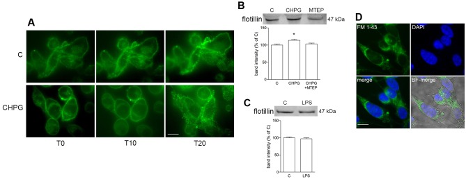
FIGURE 3.
Metabotropic glutamate 5-receptor activation increases microvesicles (MVs) release from BV2 microglial cells. BV2 cells were treated with DHPG (100 μM for 24 h) and labeled with the fluorescent dye FM1-43 (10 μM for 5 min). MVs formation following addition of Bz-ATP (100 μM) was monitored with time-lapse microscopy up to 20 min. Representative images are shown in (A). Western blot of flotillin of equal volume of MVs’ protein extracts from BV2 cells treated with CHPG (200 μM for 24 h) and MTEP (100 μM for 24 h; B), or with LPS (0.1 μg/ml for 6 h; C). Representative images of MVs shed from FM1-43-stained BV2 cells, transferred on top of SH-SY5Y cultures (D). Nuclei were stained with DAPI. Magnification = 40×(A), 63×(D). Scale bars = 20 μm (A) and 10 μm (D). ∗p < 0.05 vs. control. One-way ANOVA followed by Newman–Keuls test for significance (B) and Student’s t-test (C) were applied to detect statistically significant differences.

