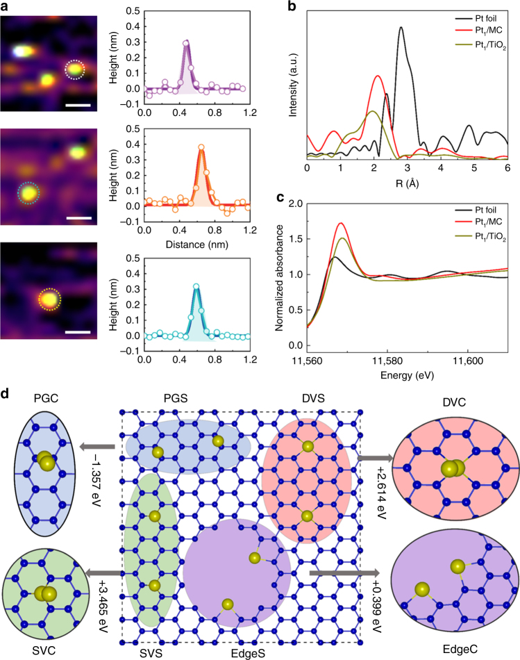
Fig. 3.
Characterizations and structure of Pt single atoms. a Typical STM images of Pt single atoms dispersed on ultrathin carbon films (constant current mode). b EXAFS spectra of bulk Pt foil and Pt single atoms absorbed on TiO2 and MC. c Normalized XANE structure spectra at the Pt L3-edge. d Structure and size distribution for Pt single atoms and Pt clusters on mesoporous carbon. The Pt single atom and clustering configurations are denoted by XS and XC, respectively, where X indicates the Pt atoms adsorbed on different defects (SVs and DVs) and edges. The yellow and blue balls represent the Pt and C atoms, respectively. (scale bar, 0.2 nm)

