Figure 4.
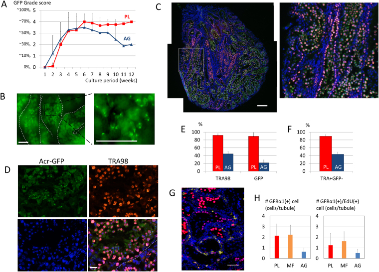
The maintenance of spermatogonia in PL device. (A) The average GFP-grade transition was compared between PL (red line) and AG (blue line) for 12 weeks. (B) At 104 culture days, the seminiferous tubules of the 2.5 dpp transgenic mouse contained GFP-positive cells including many haploid cells. (C) Immunohistological examination of the tissue cultured for 89 days at a lower magnification demonstrated GFP(+) (green) and TRA98(+)cells (red), counter-stained with Hoechst (blue). The white rectangular area is enlarged on the right. (D) Immunohistological examination at a higher magnification, GFP(+)/TRA98(+) cells were located in the inner area of the seminiferous tubule, while GFP(−)/TRA98(+) cells were near the basal membrane (broken line). Dot and crescent-like GFP condensations observed in the inner area of the seminiferous tubule represent round and elongating spermatids, respectively. (Upper left panel) (E) The rates of seminiferous tubules containing TRA98(+) and GFP(+) cells, respectively, at 12 culture weeks were compared between PL (red) and AG (blue). (F) The rates of the seminiferous tubules containing TRA98(+)/GFP(−) cells in PL (red) and AG (blue). (G) Immunohistological examination of the tissue cultured for 54 days. GFRα1(+) cells are in yellow and EdU(+) cells are in red. The nuclei were stained by Hoechst 33342 dye (blue). (H) The number of the GFRα1(+) cells and GFRα1(+)/EdU(+) cells per seminiferous tubule at 8 culture weeks were compared between PL (red), MF (orange), and AG (blue). Scale bar: 50 µm (B), 200 µm (C), 20 µm (D), 50 µm (G).

