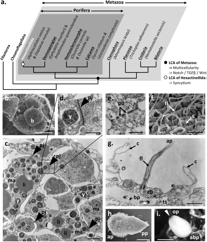
Figure 1.
Peculiar cell organization and development of glass sponges: Oopsacas minuta. (a) Phylogenetic position of glass sponges (Hexactinellida) among poriferans and metazoans. LCA: Last common ancestor. (b.,f. and h.) Scanning electron micrographs. (c.,d.,e. and g.) Transmission electron micrographs of epoxy sections. (b.) 8-cell stage embryo within maternal tissue43. (c. and d.) Details of embryo showing the fusion of macromeres and the establishment of plugged junctions. (e.) Cytoplasmic continuity between cells through plugged junction. (f. and g.) Collar bodies evidencing a clear cell polarity as shown by the apical pole, ap and the basal pole, bp. (h) Larva showing a clear anterior-posterior axis. The anterior pole and posterior pole are indicated: ap and pp respectively. (i) Adult specimen harboring a clear apico-basal axis. The oral pole (apical pole) and aboral pole (basal pole) are indicated op and abp, respectively. Abbreviations: b, blastomere; c, collar; cb, collar bodies; fl, flagellum; li, lipid inclusion; ma, macromere; nu, nucleus; os, osculum (white arrowhead); pj, plugged junction (grey arrowhead); po, pore particles; ts, trabecular syncytium; y, yolk. Scale: (b.), 43 µm; (c.), 3 µm; (d.,e. and g.), 1 µm; (f.), 4.3 µm; (h.), 50 µm; (i.), 0.5 cm.

