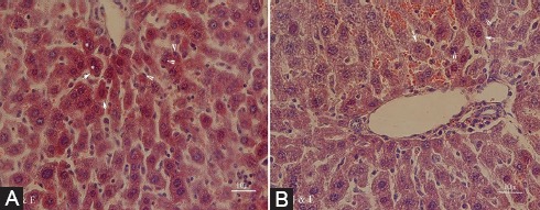
Figure 3.

Photomicrographs of the hepatic section of the DM group (A) with cytoplasmic hypereosinophilia (arrows), extensive nuclear pyknosis (arrowheads), and loss of intercellular borders in some of the hepatocytes (asterisks), and the DM plus VOO group (B) with only focal nuclear pyknosis (arrowheads) and cytoplasmic vacuolation (arrows) (stained with hematoxylin and eosin, ×400).
