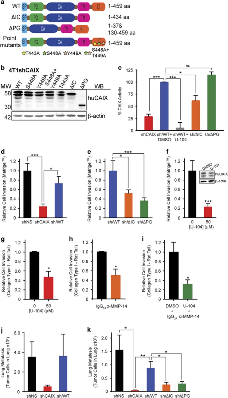
Figure 5.
CAIX regulates breast cancer cell invasion and metastasis. (a) Schematic showing the domain structure of wild type (WT) huCAIX (459 aa) and constructs lacking either the intracellular domain (ΔIC), the extracellular proteoglycan-like domain (ΔPG) or the indicated point mutations in the IC domain. SP, signal peptide; CA, catalytic domain; TM, transmembrane domain. (b) Western blot analysis of the levels of expression of the indicated huCAIX constructs by 4T1shCAIX cells cultured in hypoxia. β-actin served as a loading control. (c) Analysis of CAIX catalytic activity using the in-cell carbonic anhydrase activity assay. Assays were performed in normoxia in the presence or absence of U-104 (50 μM) as indicated. Levels of CAIX catalytic activity were normalized by using the time required to achieve 50% of the total decrease in pH. Data were normalized to the spontaneous rate of reaction in the presence of buffer alone and the activity of cells expressing WT huCAIX was set to 100%. Data show the mean±s.e.m. of technical replicates (n=3/group) and are representative of three independent experiments *P<0.05, ***P<0.001. (d) Invasion through Matrigel by the indicated 4T1 cell lines cultured in hypoxia. Data show the mean±s.e.m. of three independent experiments. *P<0.05, ***P<0.001. (e) Invasion through Matrigel by the 4T1shCAIX cell lines expressing WT, ΔIC and ΔPG variants of huCAIX and cultured in hypoxia. *P<0.05, ***P<0.001. (f) Invasion through Matrigel by 4T1 sh/WT huCAIX cells cultured in hypoxia and treated with U-104. ***P<0.001. Western blots of hypoxia-induced CAIX expression are shown as insets. For (d)– (f), data show the mean±s.e.m. of three independent experiments. (g–i) Analysis of invasion through type 1 collagen by 4T1 sh/WT huCAIX cells cultured in hypoxia and treated with (g) U-104 (50 μM), (h) anti-MMP14 antibody (20 μg/ml) or (i) a combination of anti-MMP14 antibody (20 μg/ml) and U-104 (50 μM). *P<0.05, **P<0.01. Data in (g)–(i) show the mean±s.e.m. of three independent experiments. (j) Analysis of spontaneous lung metastases formed by the indicated 4T1 cell lines following growth of orthotopic breast tumors. Data show the mean±s.e.m. n=6/group. (k) Analysis of experimental lung metastases formed by the indicated 4T1 cell lines. Mean±s.e.m. is shown. n=6/group. *P<0.05, **P<0.01. Statistical analysis was performed using ANOVA (c,d,e,k) or Student’s t-test (f,g,h).

