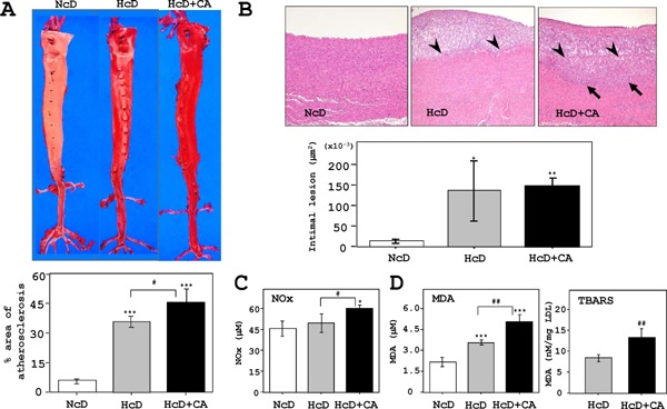
Fig. 2.

Analyses of atherosclerotic aortas.
A) The en face analysis of oil red-O-stained aortas revealed that the atherosclerotic lesion (red-stained) in HcD+CA-fed µMPs was further progressed than that in HcD-fed µMPs. B) Quantitative analyses showed that the thickening of the intimal areas of aorta was increased in HcD- and HcD+CA-fed µMPs as compared to NcD-fed µMPs. Histological examinations of the atherosclerotic lesions in HcD+CA-fed µMPs demonstrated that the thickened intimal areas predominantly consisted of accumulation of macrophages. Intriguingly, greater numbers of infiltrating foam cells were observed in the upper layer of the aortic media from HcD+CA-fed µMPs (arrows). Arrowheads denote the border between the intima and the media. C) Serum NOx levels were increased in HcD+CA-fed µMPs, in comparison to NcD- and HcD-fed µMPs after eight weeks of feeding. The NOx levels in NcD- and HcD-fed µMPs were not significantly different. D) Similarly, serum MDA levels in HcD+CA-fed µMPs were higher than those in NcD- and HcD-fed µMPs. The levels of another oxidative stress marker, TBARS, in HcD+CA-fed µMPs were increased in comparison to HcD-fed µMPs. Serum TBARS levels are expressed as nM MDA/mg LDL protein. The values represent mean ± SE. *p < 0.05, **p < 0.01, and ***p < 0.001 vs. NcD-fed µMPs; and #p < 0.05 vs. HcD-fed µMPs.
