
Fig. 4.
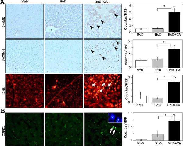
Hepatic oxidative stress markers and apoptotic activity.
A) IHC revealed that very small numbers of liver cells accumulated 4-HNE and 8-OHdG in NcD- and HcD-fed µMPs. The 4-HNE and 8-OHdG-positive cell numbers were increased in HcD+CA feeding (arrowheads). Furthermore, the numbers of DHE-positive hepatocytes in the livers of HcD+CA-fed µMPs (red-stained; arrows) were increased than in NcD- and HcD-fed µMPs after eight weeks of treatment. C= central vein. B) Significantly greater numbers of TUNEL-positive hepatocytes (green-stained; arrows) were observed in HcD+CA-fed µMPs in comparison to NcD- and HcD-fed µMPs. The apoptotic hepatocytes occasionally showed condensed chromatin and fragmented nuclei with DAPI staining (inset). The values represent mean ± SE. *p < 0.05 and **p < 0.01 vs. NcD-fed µMPs; and #p < 0.05 and ##p < 0.01 vs. HcD-fed µMPs.