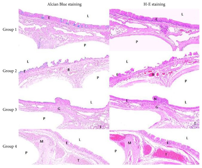
Figure 4.
Histopathological view of the tracheal graft site at the completion of the study (day 28 or death time). At day 28 after operation, the regenerated neomucosa with an epithelial lining is observed on the scaffold and tracheal luminal surface was completely covered with epithelial cells in Group 1. Marked fibroblastic proliferation, mononuclear cell infiltration, high vessel density, and epithelial thickness were observed in Groups 3 and 4. In Group 2, fibroproliferative tissue with fibrosis below the epithelium is less evident. Histopathologic examination revealed nonepithelialized epithelium (Alcian Blue and hematoxylin and eosin staining; original magnification, ×40). Group 1: no medication, Group 2: 10 mg/kg cyclosporine, Group 3: 5 mg/kg azathioprine, and Group 4: 2.5 mg/kg azathioprine plus 5 mg/kg cyclosporine. N: nonciliated cell, M: hypertrophy of mucosal smooth muscle, I: inflammatory cell, G: hyperplasia of submucosal mucous gland, L: tracheal lumen, B: blood vessels, T: trachealis muscle, E: epithelium, and P: polycaprolactone (PCL) bellows-type scaffold.

