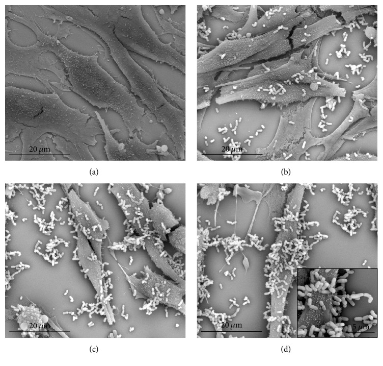
Figure 1.
Scanning electron microscope images of vaginal epithelial cells treated for 2 h with lactobacilli isolated from cocoa fermentation. (a) Untreated HMVII cells (×2,500); (b) HMVII cells treated with L. fermentum 5.2 (×2,500); (c) HMVII cells treated with L. plantarum 6.2 (×2,500); (d) HMVII cells treated with L. plantarum 7.1 (×2,500; details in ×20,000).

