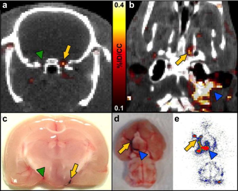
Figure 16.
PET–CT images 64Cu-FBP7 detect occlusive thrombus in both intracranial (a and b) and extracranial (b) arteries (a. arrow denotes internal carotid artery (ICA)/middle cerebral artery (MCA) with thrombus, green arrowhead denotes contralateral ICA/MCA; b. arrow denotes intracranial ICA/MCA, blue arrowhead denotes extracranial ICA). c. and d. Postmortem brains from animals injected with Evans blue-labeled clot (arrow in c denotes ICA/MCA with thrombus, green arrowhead in c denotes contralateral ICA/MCA; arrow in d denotes intracranial ICA/MCA, blue arrowhead in d denotes extracranial ICA). e. Brain autoradiography revealing high activity along the ICA and MCA (arrow denotes intracranial ICA/MCA, blue arrowhead denotes extracranial ICA). Reprinted from ref. 85 with permission. Copyright © 2014 by American Heart Association.

