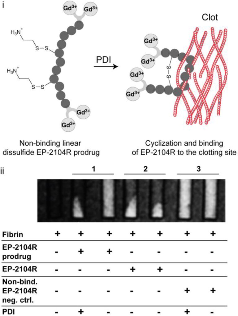
Figure 8.
PDI activation of the mixed disulfide EP-2104R prodrug. i. Schematic representation of the activation and retention approach for imaging acute thrombosis. ii. T1-weighted images of phantoms at 1.5 T after incubation under different conditions as shown in the figure and after centrifugation to pellet the clots. MR signal is only co-localized with pelleted fibrin clot if PDI is present, while without enzyme MR signal from the prodrug probe is uniformly distributed throughout the solution (column 1). Column 2 and 3 refers to positive (original EP-2104R probe) and negative (linear non-disulfide EP-2104R analogue) controls. Adapted from ref. 79 by permission of John Wiley & Sons, Inc. Copyright © 2014 by John Wiley Sons, Inc.

