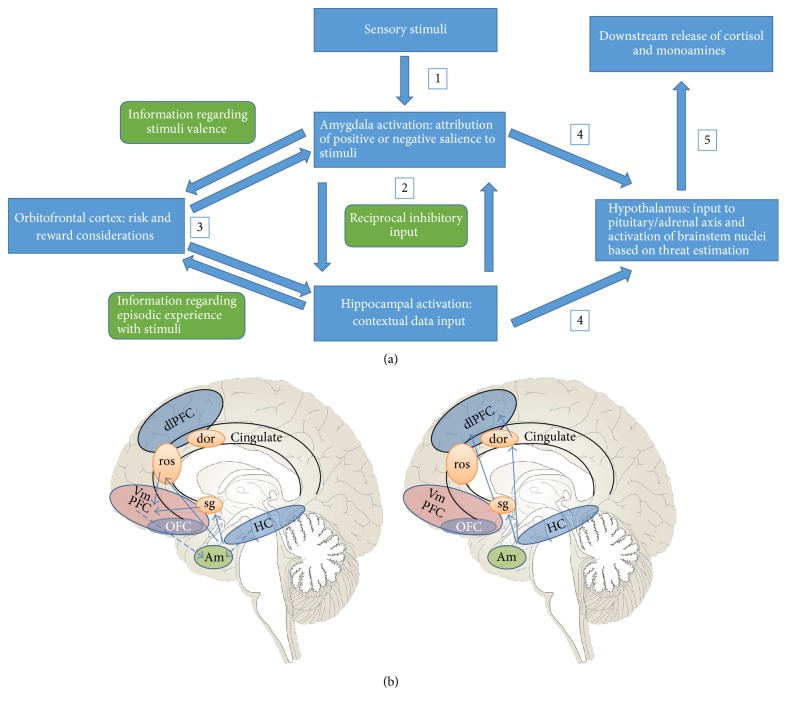
Figure 1.
Depiction of the subcortical-cortical communication which will inform whether the dlPFC or vmPFC will be preferentially activated in response to environmental stimuli. (a) shows how, as the individual perceives an element in the environment (the thalamus is not portrayed in this figure), the amygdala will be activated, and positively or negatively valenced associations will emerge based on past experience. The hippocampus will provide some level of contextual data based on episodic memory, and the OFC will weigh risk and reward considerations based on input from these two structures. Subsequent neuroendocrine responses will ensue, with the hypothalamus being more or less driven to initiate downstream cortisol release, as well as stimulating brainstem nuclei for release of monoamines, depending on the subjective sense of danger felt to be present. (b) illustrates the subsequent higher cortical level activation that will occur after this initial communication. The subcortical-cortical connection will be mediated by the ACC (either ventral or dorsal portions), which will divert activation preferentially towards the dl or vmPFC. Left panel: in instances of lower perceived environmental threat, the vmPFC is activated via portions of the sgACC and the rACC; there is greater inhibitory connection with the amygdala, thus allowing for greater top-bottom mitigation of the fear response (also aided by the inhibitory contextual hippocampal input); more robust development of the vmPFC-amygdala and hippocampus-amygdala control mechanisms can allow for more controlled responses to the environment, even when there may be potential threat, as cognitive control and contextual data will prevent excessive reactivity and stimulus generalization, permitting greater flexibility and hence more adaptive responses. The vmPFC has been shown to be hypoactive in cases of child abuse, major depressive disorder, borderline and antisocial personality disorders, and posttraumatic stress disorder, among others (refer to text for more detail). Right panel: in situations of amygdala-driven bottom-top communication, as is seen in anxiety disorders, posttraumatic stress disorder, and borderline personality disorder, portions of the sgACC and the dACC may be preferentially activated and access the dlPFC, resulting in excessive cognitive control, attempts to suppress distressing memories, and lack of attunement with one's own emotional response, given the lack of inhibitory feedback onto the amygdala; in (b), solid lines represent excitatory connections and dashed lines, inhibitory connections. dlPFC = dorsolateral prefrontal cortex; vmPFC = ventromedial prefrontal cortex; OFC = orbitofrontal cortex; Am = amygdala; HC = hippocampus; Hy = hypothalamus; sg = subgenual anterior cingulate cortex; ros = rostral anterior cingulate cortex; dACC = dorsal anterior cingulate cortex. The sgACC has connections to both the vmPFC and the dlPFC, the implications of which are described in the text. This depiction is of the medial surface of the brain; the dlPFC and portions of the OFC are located on the superolateral surface of the cerebrum, and their representations here are schematic.

