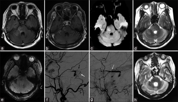
Figure 1.
Representative images of the patient. (a and d) Axial FLAIR magnetic resonance imaging and T2W showed multiple hyperintense lesions in the right cerebellum and pons. (b) This was partially enhanced by contrast. (c) DWI showed some hyperintense signals. (e) SWI showed multiple microbleeds. (f and g) Cerebral angiography showed a dAVF supplied by a distal dural branch of the occipital artery (white arrow), and (f) the petrosal branch of the middle meningeal artery (black arrow), draining into the right superior petrosal sinus (white arrow) and (g) refluxed into the perimedullary vein (small black arrows). (h) T2W showed the lesions vanished several weeks after treatment. FLAIR: Fluid attenuated inversion recovery; dAVF: Dural arteriovenous fistula; T2W: T2-weighted images; DWI: Diffusion weighted imaging; SWI: Susceptibility weighted imaging.

