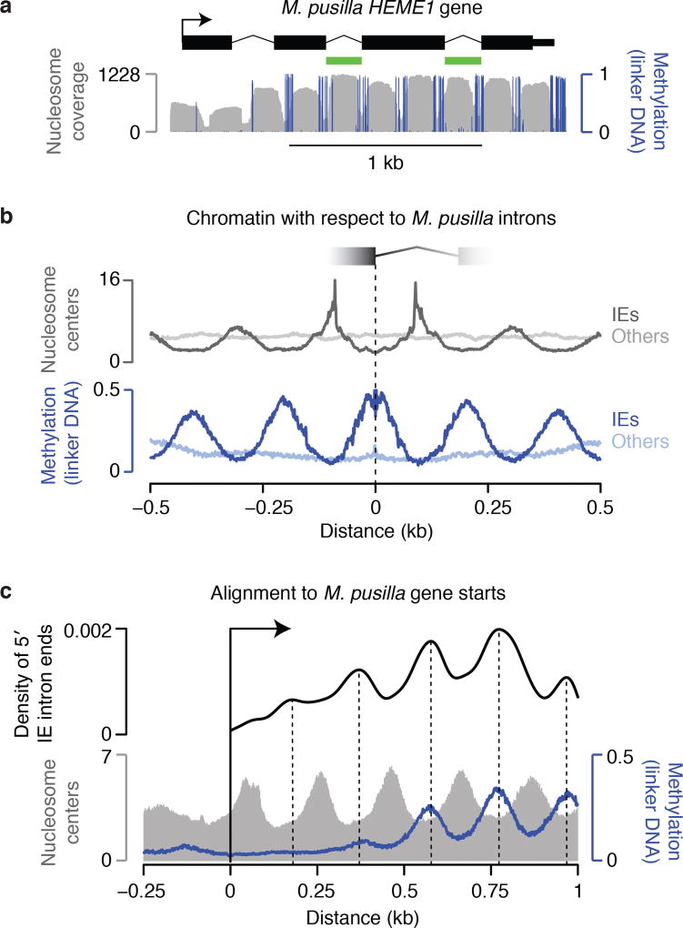
Figure 1. M. pusilla IEs insert between preexisting nucleosomes.
a, Each IE contains a nucleosome with ends in linker DNA, which is specifically marked by methylation in this organism. Validated introns and chromatin data12 are displayed. HEME1 contains 2 IEs (green). b, IE introns are generally in phase with nucleosome positions, whereas other introns are not. Chromatin maps12 are aligned to 5′ IE intron ends (dark lines) or other intron ends (light lines). c, IEs are in phase with the starts of genes, indicating insertion between preexisting nucleosomes. Chromatin maps12 and 5′ IE ends are aligned to gene starts. A kernel density estimate of IE ends is shown with peaks marked.

